What is an Eyelid Tumour?
An eyelid tumour is when an abnormal mass of new tissue forms on the upper or lower eyelid. They can either be benign, which means they do not invade other tissues, or malignant, which denotes an ability to infiltrate tissues surrounding the lesion and to spread to other parts of the body – or to “metastasize”.
The eyelid is a very common site for tumours as it is sun exposed and the tissues are histologically different to the rest of the body. Tumours can spread rapidly and often the roots of the tumour are much larger than is visible on the surface of the skin. For this reason, it is important to be assessed as soon as a new lesion is noticed, to prevent spread throughout the eyelid or to the rest of the body.
What Types Of Eyelid Tumours Are There?
Multiple types of benign lesions including cysts, skin tags and naevi are commonly seen on the eyelids. It can sometimes be difficult to differentiate between benign and malignant lesions on the examination findings and history alone. We often recommend biopsy to exclude a malignant lesion, as left untreated can lead to serious consequences.
The most common types of malignant eyelid tumours are:
- Basal Cell Carcinoma - Basal cell carcinomas or ‘BCCs” are the commonest form of eyelid tumour and usually present as raised, pearly lesions on the eyelid margin. These are unlike other forms of cancer in the fact that they do not spread to other parts of the body. They can, however, grow quite large locally and invade other tissues. This type of tumour is more common in fair-skinned people and usually affects older individuals although can affect younger people too.
- Squamous Cell Carcinoma – This is an aggressive form of skin cancer which forms from squamous cells which are the top layer of the skin. These can spread rapidly to other parts of the body, although when caught early, most SCCs are curable. SCCs have a wide range of presentation from scaly, pink skin to a keratinised cutaneous horn.
- Sebaceous Gland Carcinoma – These very rare but aggressive tumours grow from the sebaceous glands found in the eyelids, from which oil is produced to keep the eye moist. These tumours are often misdiagnosed as a chalazion or blepharitis and therefore it is essential to be reviewed if you have a chalazion for many months or unilateral blepharitis.
What Are The Signs and Symptoms of Eyelid Tumours?
Most eyelid tumours are asymptomatic in their early stages. Signs can vary but are commonly characterised by skin changes such as raised bumps, which may or may not be pigmented. Tumours in their advanced stage can ulcerate or display scaling and crusting. The loss of eyelashes is an indicator of a malignant tumour.
If the cancer has started to spread, patients may experience swelling of the lymph nodes around the ear and under the chin. In later stages, patients may present with orbital pain, reduced vision, double vision or displacement of the eye.
Symptoms of eyelid tumours can vary, but are commonly characterised by skin changes. Tumours in their advanced stage can ulcerate or display scaling and crusting, while earlier signs include lumps, lesions or bumps. If the cancer has started to spread, patients may experience swelling of the lymph nodes around the ear and under the chin.
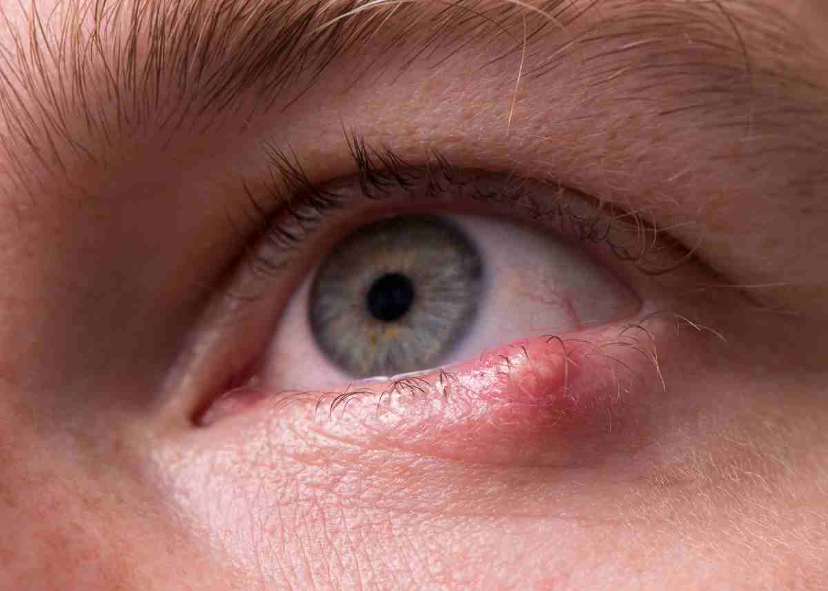
Basal cell carcinoma brings about a painless hard lump, usually in the lower eyelid, that grows to break down deeper tissue. The loss of eyelashes is an indicator of the tumour becoming malignant.
Symptoms of Squamous cell carcinoma sometimes appear as a non-healing ulcer on the lower eyelid with hard raised edges, featuring redness, crusting or bleeding. Patience may experience decreased vision if the cancer spreads behind the eye. Sebaceous gland carcinoma commonly displays a small but firm red or yellow lump on the eyelid. This may gradually increase in size and cause irritation to the eye.
What Are The Causes of Eyelid Tumours?
In most cases the cause of a tumour is not known, but there are clear risk factors which have been proven to increase the risk of them developing. Environmental factors such as excessive sun exposure and consequent skin damage, especially in fair skin skinned individuals are a major risk factor. Genetics appear to play a role and a suppressed immune system, due to medication or disease can increase the risk of tumours developing. There are pre-malignant lesions such as Bowens Disease, actinic keratosis, and keratoacanthomas which can develop into malignant lesions if left untreated.
How Can You Prevent Eyelid Tumours?
Sun protection is paramount in preventing all forms of skin cancers. To protect the eyes, sunglasses and a hat should be worn outside.
Any new lesions that develop should be examined for signs of malignancy before they can progress. Although benign tumours don’t require active treatment, they should be frequently monitored for changes. If they are cosmetically challenging or posing a level of discomfort, they can be surgically removed. Early detection of malignant tumours is crucial, to prevent local spread and distant metastases.
What Are The Treatments For An Eyelid Tumour?
There are a wide range of treatment options available at The Daniel Ezra Clinic. Your ophthalmologist will advise which is the best course of treatment for you:
- Topical chemotherapy – A chemotherapy ointment that is directly applied to the tumour
- Cryotherapy – An ice-cold metal probe is used to destroy the cancer cells
- Laser Treatment – A focused beam of light is used to destroy the cancer cells
- Radiation – The tumour is exposed to high-powered X-ray beams to kill its cancer cells
- Surgery – The tumour is surgically removed by a MOHS or oculoplastic surgeon. After confirming that all cancerous cells have been removed, the patient can then move on to eyelid reconstruction during a separate operation.
- Systemic chemotherapy – Patients with advanced disease may be treated with chemotherapy drugs to treat the tumour and any metastases.
What is the process for tumour diagnosis and management?
The first step in your treatment will be to establish an accurate diagnosis through a detailed history and examination. Many benign lesions have typical features and you may be reassured that there is no cause for concern. Often a biopsy is required for a definitive diagnosis which is performed as a day case procedure under local anaesthetic. The area is numbed with a small injection and a sample of the lesion is sent to the laboratory for histopathology. The results of the biopsy usually take around 1 week.
Your ophthalmologist will review the results of the biopsy and explain the treatment options to you. If the biopsy reports a BCC, an excision and reconstruction will be planned. With all other forms of skin cancer, further investigations including blood tests, scans and ultrasounds will be arranged in addition to the excision and reconstruction.
At the Ezra Clinic, tumours can be excised with a margin of a few millimetres of normal tissue to ensure clearance of the surgery or by MOHS surgery. A reconstruction of the lid will be planned following excision of the tumour.
What is MOHS surgery?
MOHS micrographic surgery is considered the most effective technique for treating BCCs and SCCs. The procedure is performed by a dermatologist who is able to both surgically excise the tumour and examine it under a microscopy on the same day.
Mohs is performed in stages; the dermatologist excises the area they believe to be involved with tumour and examines this under the microscope. If the tumour has spread to involve one of the surgical margins, a second stage will be performed to excise further tissue in this area. The process is repeated until the tumour is removed in total.
MOHS surgery has two main advantages. Firstly, it has a very high cure rate as the dermatologist can be confident that the tumour has been completely excised. Secondly, Mohs allows you to keep as much healthy skin as possible as the dermatologist only removes the affected skin. This is especially important on the eyelid when you need to preserve as much normal tissue as possible.
What is a reconstruction?
Skin tumours have roots which often extend much larger than is visible on the surface of the skin. Following excision of a tumour, patients may have a defect much larger than expected from a relatively small skin lesion. In some cases, the skin edges may be pulled together. In larger defects there are a variety of techniques that can be used including skin flaps and grafts from other parts of the body. Eyelids have an excellent blood supply and often heal very well following reconstruction.
What are the complications that come with eyelid tumours?
Overall, eyelid tumours are fairly common, and have low rates of spread. Nonetheless, tumours can pose a high risk of damage to nearby eye structures which could lead to serious complications such as dry eyes, incomplete eye closure, double vision and blindness, and if left untreated, certain cancers may well spread to the rest of the body.
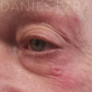
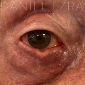
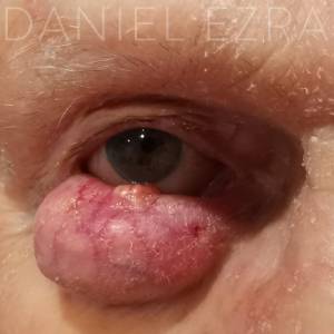
Examples of Eyelid Tumours and Lump on Bottom Eyelid